


30.05.2022
Mathematical model to mould an implant to the bone

AGH UST scholars develop a numerical model that allows them to simulate the behaviour of bone tissue under mechanical loading, photo from Dreamstime
Bone tissue can change its structure multiple times during a lifetime, adapting to various loadings that our bodies may be exposed to. Implants can disrupt this natural process. Therefore, AGH UST scholars develop a numerical model that allows them to simulate the behaviour of bone tissue under mechanical loading. In the future, it may prove useful in designing tailor-made endoprostheses.
Bone tissue is made of compact bone and cancellous bone. The former makes up the outer layer and the shaft of long bones; the latter can be found in the epiphyses and is characterised by its trabecular structure. Julius Wolff, a German surgeon born in Märkisch Friedland (currently Polish Mirosławiec), in his 1892 monograph titled Das Gesetz der Transformation der Knochen [The Law of Bone Remodeling], popularised the idea that the arrangement of struts (trabeculae) reflects the direction of major loadings on the bone. Moreover, he observed that every change of form and function of a bone entails specific changes in its internal architecture as well as external conformation of the shape according to mathematical laws. This claim was later named “Wolff’s law”, and its creator was considered one of the forefathers of orthopaedics as a separate medical discipline.
However, Wolff has never proposed any mathematical rules himself. It was an American orthopaedist and surgeon, Harold Frost, who, in a series of articles printed in the 1960s, demonstrated the premises of Mechanostat. The theory implies that the reshaping of the bone architecture is due to mechanical loading. If the loading oscillates within certain boundaries, the biological balance is maintained. However, when the upper limit is exceeded, the tissue grows. And when the lower limit is overstepped, an inverse process occurs. This is why, as a result of training, the legs of long-distance runners adapt to extreme strains and the bones of bedridden patients resorb and require rehabilitation during recovery. This occurs due to the change in bone density or the reorientation of the trabeculae.
 An image of the arrangement of taberculae in a fragment of a thigh bone and its computer model, image by the Laboratory of Micro- and Nanotomography
An in-depth understanding of this mechanism carries meaning for bone grafting, when, for example, there’s a need to replenish bone loss resulting from cancer or hip replacement surgery. ‘On Earth, with the typical loading that affects us, our skeletons and muscles remain in balance. By inserting an implant into our bodies, we disrupt this natural distribution of stresses and our bone tissue must adapt to the new conditions. However, if the implant wasn’t properly placed or its shape wasn’t optimal, some fragments of bone tissue may be exposed to lesser or greater loading than in the case of healthy bone. This, in turn, can lead to bone atrophy within the area of implantation. This may loosen the implant and, as a consequence, lead to its removal’, explains Sebastian Wroński, an AGH UST Professor at the Faculty of Physics and Applied Computer Science, who specialises in materials science.
Fully anisotropic model for bone tissue remodelling
AGH UST Professor Wroński, as part of a project he leads, similarly to other teams working on the issue worldwide, develops a numerical model for bone tissue remodelling under mechanical loading. In his work, the scholar focuses on the femur, which is the longest bone in the human body and allows it to remain and move upright. Its head makes up an element of the hip joint, where it sustains the loading generated by body weight transferred from the pelvis. On the outside, it is made of compact bone, on the inside – cancellous bone with a trabecular structure.
The model developed by Professor Wroński, to be able to mirror the loading on the thigh bone as accurately as possible, accounts for not only the remaining bones of the lower extremity, but also its most important muscles. Modelling an entire limb with the muscular system allows the scientist to simulate dynamic loading related to various positions of the leg, e.g., during walking. Furthermore, as the researcher claims, the model developed at the AGH UST has at least two qualities that distinguish it from the competition.
To begin with, it completely accounts for the anisotropic properties of bone tissue. ‘The spatial network of trabeculae that make up the cancellous bone, may be described graphically as a very extensive lattice that exhibits a certain preference in terms of the orientation of the trabeculae. When we apply force to such a spatial structure, its behaviour will depend on the direction of this force. The elasticity of this material can vary substantially depending on the direction in which you study it’, Professor Wroński explains.
The other advantage of the solution proposed by the AGH UST scholar is the implementation of a mechanism that is responsible for the change in the spatial structure of the trabeculae during the remodelling of bone tissue. ‘In the skeletal system, there are two types of cells. Osteoclasts to resorb the bone and osteoblasts to build it. When balanced, the density of the latter remains unchanged because the same amount of cells is resorbing and growing bone. However, the latter can do that in different directions! Then, despite the state of balance, the trabeculae re-orientate. Taking this effect into account is one of the greatest pros of this model’, the scholar highlights.
Ultra-high-resolution tomography
Developing such a precise model would not have been possible without the Laboratory of Micro- and Nanotomography (LMiNT), established at the AGH UST Faculty of Physics and Applied Computer Science in 2012. The unit was created on the initiative of Professor Wroński and another AGH UST Professor, Jacek Tarasiuk, who is currently its head. ‘A technology that captivated me to the extent that after 15 years of work, after having obtained my postdoctoral qualifications, I walked away from my previous domain to get involved in something I thought was extremely attractive’, recalls Professor Tarasiuk, talking about micro- and nanotomography.
The LMiNT, as one of the few laboratories in Poland, has a high-resolution CT scanner that allows scientists to investigate 3D structures of objects with a resolution of 300 nm. In addition, it also has a miniature universal testing machine, which allows researchers to observe “live” how the microstructure of a material reacts to the given load. Due to its unique capabilities, the machine is used on a daily basis to carry out numerous tests for the needs of science and industry. Among other things, scientists test rocks and their fractures, ceramic materials, composites, polymer substitutes for bone tissue (scaffolds), heart valves and the calcification thereof, meteorites, arctic lichen, teeth with various types of fillings, polychrome, welds, and adhesive joints.
However, from its inception, the focus of the creators of LMiNT revolved around human bones. ‘We carry out research that will make it easier to design implants. To create such models, we need to experimentally verify the structure of a bone and its reaction to loads. In our lab, we try to find a connection between the structure and internal architecture of a bone and its reaction to mechanical loading. Subsequently, we use this knowledge to build computer models’, the scientists explained 10 years ago, when the laboratory had just launched.
Successes of AGH UST alumni
The approach that is based on using computed tomography for, on the one hand, obtaining output data for mathematical modelling and, on the other, the verification of proposed models has quickly born fruit. A testimony of that is the successes of our graduates who, still being students, developed their knowledge in various projects in this domain:
- Jakub Kamiński, MSc Eng – first prize in a university grant competition, an honourable mention in the competition organised by the Polish Biomedical Engineering Association, and the President of Krakow award for his thesis on modelling the structure and mechanical properties of cancellous bone (2013)
- Bartłomiej Mitka, MSc Eng – second prize in the competition organised by the Polish Biomedical Engineering Association and the President of Krakow award for his thesis on computer simulation of bone tissue remodelling with the use of the finite element method (2014)
- Paweł Miry, MSc Eng – an honourable mention in the competition organised by the Polish Biomedical Engineering Association for his thesis on the anisotropic model of bone tissue remodelling under mechanical loading (2016)
Tailor-made implants for patients
In the future, solutions similar to Professor Wroński’s model may be the reason implants will be prepared with the anatomical architecture of individual patients in mind. The growing omnipresence of 3D printing and, consequently, its dropping costs, may result in implants designed on the basis of computer simulations being produced directly in hospitals. To get there, we need to collect appropriate data for our mathematical models. This is where it gets bumpy because the CT scanners that are currently used in hospitals have far worse parameters compared to the LMiNT equipment. ‘When we perform a CT scan in a hospital, the resolution is not satisfactory enough to precisely describe the internal architecture of a bone. We can only see areas with higher or lower density, but we cannot see the spatial trabecular structure clearly enough’, Professor Wroński describes.
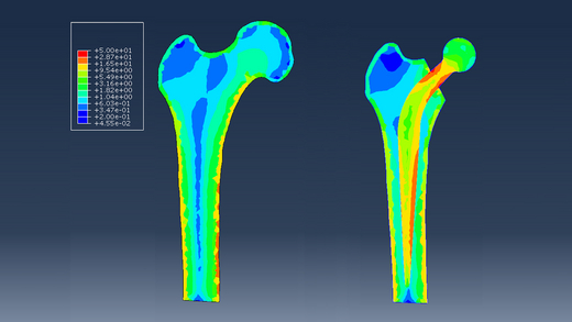 Distribution of stress in a healthy bone and a bone with an implant, image by the Laboratory of Micro- and Nanotomography
However, the AGH UST scientists want to take a leap into the future by tweaking ready-to-implement solutions, when precise tomography will not just be within the reach of specialised academic laboratories. ‘The model is operational, tested, and the concept is published. The next step is to apply the model to a specific case. We are thinking of establishing cooperation with a group of doctors who want to research this field. Whether the investigation shall concern hip implants or bone damaged as a result of cancer depends on the group with which we’ll cooperate’, predicts Professor Wroński.
The project titled Development of bone adaptation model under external loads received funding from the “SONATA BIS 7” programme of the National Science Centre.
All rights reserved © 2021 AGH University of Science and Technology